True 3D image access for the Clinician
Surgeons and treating clinicians require better 3D imaging and interactive capabilities than is currently available.
TRUE LIFE ANATOMY provides that technology.


The Need
To optimize patient care delivery, the treating clinician needs good diagnostic imaging and interactive 3D image access to assess the patient’s patho-anatomy with true and accurate measurements, and full 3D perspective.

The Problem
Current volume rendering 3D imaging programs can only deliver 2D screen captures to the clinician.
The clinician cannot truly manipulate the image, in a cost effective platform.

The Solution
TRUE LIFE ANATOMY delivers truly interactive 3D image capability to the clinician within a cost effective and accessible format with the capacity for virtual surgery, arthroplasty templating and patient instruction.
True Life Anatomy
Advanced interactive 3D imaging
TLA provides true 3D interactive capability within a cost effective and accessible format with the capacity for virtual surgery, arthroplasty templating and patient instruction – this solution is market desirable, economically attractive and a sound investment to complement existing imaging capabilities.

Current situation and
limitations in Technology
Most current Radiology based 3D CT imaging programs use volume rendering (VR) technology. While this provides outstanding direct visualization of the 3D data set, and can effectively display soft tissues, there are limitations in sharing and modifying this image.
The value to the clinician is limited as the parts of the image cannot be individually manipulated or moved. This is because VR only displays a 2D projection (ray casting) of the individual data points, which are given variable colour and transparency to simulate a 3D image – a type of hologram.
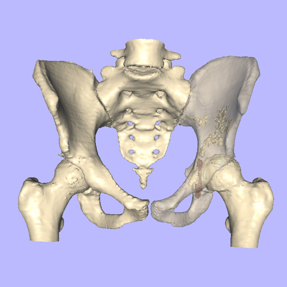
TRUE LIFE ANATOMY provides an interactive surface rendered 3D model that can be manipulated, exported and saved as a true 3D object.
Who can benefit from True Life Anatomy
3D imaging Technology?

Clinicians
Through better planning, diagnostic and treatment capabilities for clinicians.

Radiologists
Through better, faster, and a more cost efficient service to referring clinicians.

Patients
The advanced technology TLA provides, translates into better outcomes for patients.
WHO ELSE BENEFITS?
Prosthesis vendors can provide 3D model of implants to the surgeon in a protected and secure environment to allow 3D templating and planning without the significant on-costs of graphics artists and technicians. TLA utilizes existing funding pathways and resources to provide a cost-effective capability for diagnosis, planning and virtual surgery.
BOTTOM LINE – WHO PAYS?
TLA has a UNIQUE DELIVERY PATHWAY to enhance the patient care axis that uses existing skills of users, utilizes current funding resources and avoids the increased technician and 3D model printing costs. For a modest investment, the imaging provider can incorporate the enhanced 3D imaging capability and these can be can be shared with the referring clinician via a PACS / network or specially designed high-capacity USB card. The USB data card contains all the required applications and 3D manipulation tools.
The referring clinician can then perform the virtual surgery and templating incorporating 3D prosthesis data shared via a secure portal and format. This empowers the radiologist, the treating clinician and the patient within an existing funding environment.